
Last Updated: November 1, 2026
Quick Summary
Glaucoma screening for seniors is recommended every 1-2 years for those 65 and older with no additional risk factors, and annually or more often if you have a family history of glaucoma, diabetes, or high blood pressure. Screening combines eye pressure measurement, visual field testing, and OCT imaging to catch optic nerve damage before symptoms appear.
- Glaucoma typically produces no symptoms until significant optic nerve damage has already occurred.
- Age is the strongest risk factor, with odds climbing each decade after 60.
- A comprehensive screening exam is generally comfortable and usually takes less than an hour.
- An abnormal screening result is common and usually not glaucoma, so follow-up timing matters.
- Bring a medication list and arrange a ride if dilation is likely to blur your vision.
Table of Contents
- Why Glaucoma Screening for Seniors Matters More Than Ever
- What Glaucoma Is and Why It Steals Sight Quietly
- Glaucoma Symptoms vs Normal Vision Loss
- Age and Other Risk Factors That Raise Your Odds
- What Happens During a Comprehensive Eye Examination
- How an OCT Scan for Glaucoma Sees What Pressure Alone Misses
- OHIP Eye Exam Coverage for Seniors: What Is Covered and What Is Not
- How Often Should Seniors Be Screened? Evidence-Based Intervals
- Screening Versus Diagnosis: What Your Results Actually Mean
- Preparing for Your Appointment and Making the Visit Comfortable
- Frequently Asked Questions
Why Glaucoma Screening for Seniors Matters More Than Ever
Glaucoma screening for seniors is one of the most important ways to catch a sight-threatening disease before it causes noticeable symptoms. Glaucoma is a leading cause of irreversible blindness, and the risk climbs with age. Because the disease often develops silently, many people have lost vision before they notice a problem.
Here’s the uncomfortable truth most guides skip: you cannot feel glaucoma. There is no ache, no redness, no warning twinge. By the time a senior notices a missing patch of peripheral vision, optic nerve damage is already permanent. That is why regular screening matters more than symptom-watching.
This guide walks through what glaucoma is, who is at risk, what happens during a comprehensive eye examination, how OHIP coverage generally works for seniors, and how often screening is typically recommended.
What Glaucoma Is and Why It Steals Sight Quietly
Glaucoma is a group of eye diseases that damage the optic nerve, the bundle of nerve fibers connecting your eye to your brain, usually in connection with elevated pressure inside the eye. That damage narrows the field of vision over time, starting at the edges where most people aren’t looking.
The word “silently” is doing a lot of work here. Open-angle glaucoma, the most common form, produces no early symptoms at all. Vision stays sharp in the center while peripheral vision erodes in patches. Many people compensate by turning their heads slightly, never realizing they are working around blind spots.
The optic nerve and eye pressure connection
Eye pressure, or intraocular pressure, is maintained by fluid that circulates inside the eye. When that fluid drains too slowly, pressure rises and presses on the optic nerve. High pressure is not the whole story, since some people develop glaucoma at normal pressure, but it remains the biggest single risk factor clinicians track. The goal of screening is to catch pressure problems and nerve changes early, when treatment can slow the damage. Treatment generally cannot reverse vision loss, so early detection is important.
Glaucoma Symptoms vs Normal Vision Loss
Normal age-related vision change is gradual and predictable: you need more light to read, small print blurs, and glare becomes annoying. Glaucoma symptoms look nothing like that. The disease typically produces no symptoms until significant optic nerve damage has occurred, which is precisely why screening exists.
When symptoms do appear, they tend to be subtle at first. Watch out for these warning signs, which are not normal aging:
- Loss of peripheral vision, often described as tunnel vision
- Patchy blind spots in side or central vision
- Difficulty adjusting to dark rooms
- Blurred vision that comes and goes
- In acute angle-closure glaucoma, sudden severe eye pain, headache, nausea, and halos around lights (a medical emergency)
The trap is mistaking early peripheral loss for clumsiness or “just getting older.” If you bump into objects on one side or miss things at the edge of your vision, that deserves an exam, not a shrug.
Age and Other Risk Factors That Raise Your Odds
Age is the strongest risk factor for glaucoma, and the odds climb with each decade after 60. But age is not the only lever. Family history matters enormously: having a parent or sibling with glaucoma raises your risk substantially, which is why we ask every senior about relatives with the disease. If that describes you, understanding whether glaucoma is hereditary is a smart first step.
Other established risk factors include:
- Elevated eye pressure, the leading modifiable factor
- Family history of glaucoma
- African, Hispanic, or Asian ancestry, which carries higher risk for certain forms
- Diabetes and high blood pressure
- Long-term steroid use, including some eye drops
- Thin corneas and high nearsightedness
- Prior eye injury or surgery
Knowing your risk profile changes how often you should be screened. A 65-year-old with no risk factors and a 65-year-old with a parent who has glaucoma should not follow the same schedule.
What Happens During a Comprehensive Eye Examination
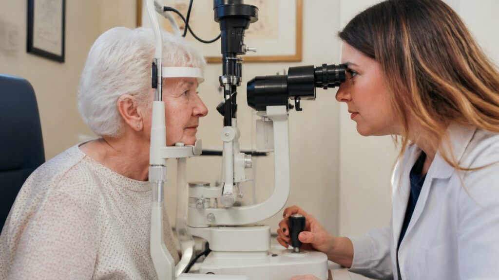
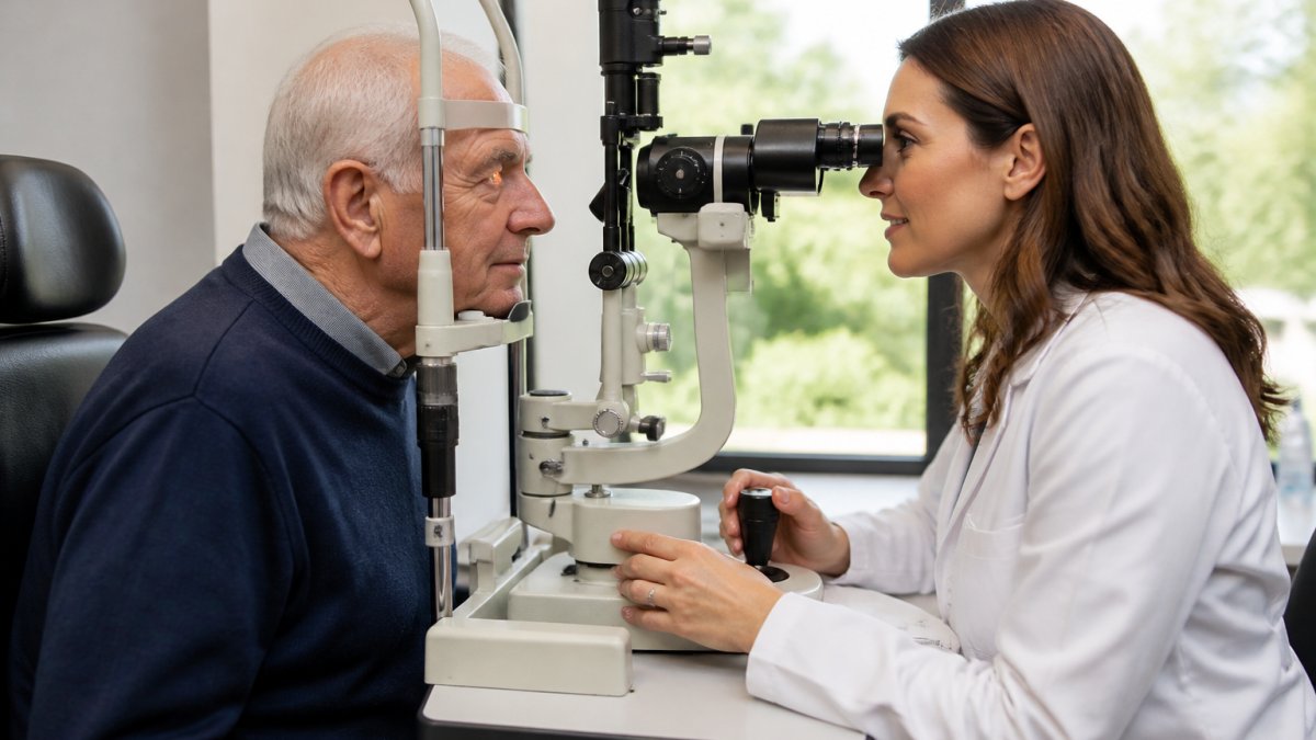
A comprehensive eye examination for glaucoma screening is generally comfortable and usually takes less than an hour. Learn the various steps taken during a comprehensive eye exam before you book. It combines several tests, each checking a different piece of the picture, so no single measurement has to carry the whole diagnosis. Here is what a senior patient can expect.
Eye pressure measurement and visual field testing
Tonometry measures intraocular pressure. A quick puff of air or a gentle probe touched to the eye’s surface gives a pressure reading in seconds. Visual field testing maps your peripheral field of vision: you look straight ahead and signal when you see lights appear at the edges. This test reveals blind spots you may not have noticed. Together, these two checks form the backbone of routine glaucoma screening, and both are quick enough that even anxious patients manage them comfortably.
How an OCT Scan for Glaucoma Sees What Pressure Alone Misses
An OCT scan for glaucoma uses light waves to build detailed cross-sectional images of the retina and optic nerve, letting your optometrist measure the nerve fiber layer thickness directly. This matters because pressure-only screening has a real blind spot: some people have high pressure without nerve damage, and others develop glaucoma at normal pressure. OCT catches what tonometry alone cannot.
OCT scans are generally non-invasive, and whether dilation is needed depends on the individual exam. By comparing your optic nerve measurements over time, your eye-care provider can detect thinning before it shows up on a visual field test. Learning what an OCT eye scan checks for helps seniors understand why this imaging has become a standard part of modern glaucoma screening.
OHIP Eye Exam Coverage for Seniors: What Is Covered and What Is Not
OHIP eye exam coverage for seniors is set by the province and can change, so check the current terms with the official source before your visit. Coverage details and any changes are set by the provincial government, so confirm current terms through Ontario’s OHIP eye care information before your visit.
Some add-on tests and follow-up visits may not be fully covered. Because coverage rules can change, ask your provider what is included before your appointment.
How Often Should Seniors Be Screened? Evidence-Based Intervals
Screening intervals are not arbitrary. They are set by balancing the slow, silent progression of glaucoma against the cost, inconvenience, and low risk of repeated testing. For seniors, the practical question is not whether to be screened but how often, and the answer depends on age, risk profile, and prior findings.
A general framework for screening intervals often looks like this (confirm with your eye-care professional):
| Risk profile | Suggested screening interval | Rationale |
|---|---|---|
| Age 65+, no additional risk factors | Every 1-2 years | Baseline risk rises with age; annual or biennial exams catch change without over-testing |
| Age 65+, family history of glaucoma | Annually, sometimes every 6-12 months | First-degree relatives carry substantially higher risk, and progression can be faster |
| Diabetes or high blood pressure | Annually | Vascular risk factors compound optic nerve vulnerability |
| Elevated eye pressure or borderline optic nerve findings | Every 6-12 months, as directed | Suspicious findings need closer monitoring to distinguish stable from progressive change |
| Established glaucoma diagnosis | Monitoring schedule set by your eye-care provider, often every 3-6 months initially | Treatment response and progression dictate follow-up frequency |
Recommendations on exam frequency vary, but eye-care professionals generally consider age, family history, and other risk factors when setting the interval. Your optometrist can advise what is appropriate for you. Find out how often you should get your eyes examined based on age, health factors, and more.
Three principles explain the intervals:
- Glaucoma progression is measured in months and years, not days. Intervals are short enough to detect meaningful change but long enough to avoid unnecessary repeat testing.
- Risk factors are cumulative. Age plus family history plus diabetes is not three separate risks; it is a compounded risk that shortens the appropriate interval.
- Prior findings reset the clock. A borderline optic nerve or a visual field defect moves a patient from routine screening to active monitoring, regardless of age.
If you are unsure where you fall, talk with your optometrist about how often you should have an eye exam based on your age and health factors. The key is to treat the interval as a clinical decision, not a fixed rule, and to revisit it whenever your health or family history changes.
Screening Versus Diagnosis: What Your Results Actually Mean
Screening and diagnosis are different stages of the same journey, and understanding the difference removes much of the anxiety around an abnormal result. Screening is a broad check that flags suspicion. Diagnosis is a focused evaluation that confirms or rules out disease. Most seniors will never move past screening; some will need follow-up; a smaller number will be diagnosed and begin treatment.
Here is how results typically unfold:
Normal screening result. Your eye pressure, optic nerve appearance, and visual field are within expected ranges. No further testing is needed until your next scheduled exam. Normal results do not mean you can skip future screening, because glaucoma can develop between visits.
Suspicious screening result. One or more findings fall outside the normal range: pressure is higher than expected, the optic nerve looks asymmetric, or a visual field test shows a possible defect. This is not a diagnosis. It is a signal to repeat testing, often with OCT imaging and a second visual field test, to see whether the finding is real and stable or a one-time variation. Many suspicious findings turn out to be normal after repeat testing.
Confirmed diagnosis. If repeat testing and imaging confirm optic nerve damage consistent with glaucoma, your eye-care provider will discuss treatment. First-line treatment is usually pressure-lowering eye drops, with laser therapy or surgery considered when drops are insufficient or impractical. Diagnosis also triggers a monitoring schedule, often every 3-6 months initially, to confirm that treatment is working and that vision is stable.
Two practical points matter for seniors:
- An abnormal screening result is common and usually not glaucoma. Elevated pressure alone, without nerve damage, is not glaucoma. Many people with higher pressure never develop the disease.
- Follow-up timing matters. If you are asked to return in three or six months, that interval is clinical, not administrative. Delaying follow-up can allow progression to go undetected.
If you are diagnosed, treatment typically begins with pressure-lowering eye drops, and monitoring continues for life to protect remaining vision and independence. The goal is not to reverse damage but to slow it enough that your functional vision lasts as long as you do.
Preparing for Your Appointment and Making the Visit Comfortable
A little preparation makes a senior eye exam smoother and more useful. Bring a list of all medications, including eye drops and steroids, plus the names of any relatives with glaucoma. Arrange a ride if dilation is likely, since your vision may stay blurry for a few hours. Write down your questions in advance so nothing slips your mind in the chair.
Accessibility matters. Alcona Eye Care offers multilingual services in English, Spanish, French, Portuguese, and Italian, and our Crossroads Plaza location offers free parking. If a rushed appointment has left you feeling like a number in the past, tell us your concerns up front so your visit can be planned around your questions.
Glaucoma gives no warning, but screening does. If you or a parent is due for a senior eye exam, don’t wait for symptoms that may never come. Alcona Eye Care provides comprehensive eye care services for patients of all ages in Innisfil, including eye exams and specialized care, delivered by a multilingual team. Book a comprehensive adult and senior eye exam and get a clear picture of your eye health. Contact Alcona Eye Care to arrange an appointment.
Frequently Asked Questions
What is the first test performed during glaucoma screening?
Most exams begin with a check of your eye pressure, called tonometry. The optometrist may use a quick puff of air or numbing drops with a small probe. Pressure alone does not confirm or rule out glaucoma, so it is paired with an optic nerve evaluation and often a visual field test.
Are eye exams for glaucoma covered by OHIP for seniors?
OHIP eye exam coverage for seniors is set by the province and can change, so check the current terms with the official source. Ask your optometrist which tests fall under the covered exam and which are optional add-ons. Bring your health card and a list of medications to every visit.
How often should OCT be done for glaucoma?
There is no single schedule. How often an OCT scan is repeated depends on the individual, and your optometrist will advise on an appropriate interval for you.
What is the difference between normal vision changes and glaucoma symptoms?
Normal age-related vision loss tends to be gradual and affects reading or fine detail first, often with blurriness in the center. Glaucoma symptoms vs normal vision loss is a key distinction: glaucoma typically starts with peripheral vision loss and blind spots you may not notice until damage is advanced. Any new loss of side vision or halos around lights deserves a prompt exam.
Can I get a free eye test if my grandmother had glaucoma?
A family history of glaucoma raises your risk, but it does not automatically qualify you for a free exam. Coverage rules are set by the province and can change, so check the official source or ask your optometrist about fees and any coverage that may apply to you.
Is OCT testing necessary for glaucoma diagnosis?
OCT is not the only test, but it adds detail that pressure measurement and visual field testing cannot show. It produces cross-sectional images of the optic nerve and retina, helping your optometrist detect thinning before vision loss becomes obvious. Many practices now use OCT alongside a comprehensive eye examination to confirm findings and monitor change over time.